Decision making for retreatment of failures in dental medicine - DVD Compendium
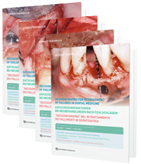

Decision making for retreatment of failures in dental medicine - DVD Compendium
€198,00
€50,00
Tabanella, Giorgio
Decision making for retreatment of failures in dental medicine
DVD Compendium
1st Edition 2018
Video DVD
4 DVDs and detailed Engl. booklets in a hardcover, NTSC/PAL
Languages: English, German, Italian
Subject: Oral Surgery
Title-No.: 3800
ISBN 978-1-78698-006-9
Quintessence Publishing, Germany
Increasingly, modern periodontology and implantology face mistreatments, complications, failures, and even iatrogenic damages. It is therefore deemed valuable for colleagues and patients to share protocols, techniques, instruments, materials, and innovations developed to retreat these clinical cases with no room for error. All clinical cases described have only one parameter in common: a medical mistake.
Retreating these "special patients" requires passion, ethics, and the willpower to help patients who present with very high expectations and an exacting psychological makeup. Variables such as periodontal biotype, biologic and anatomic potential for tissue regeneration, and the nature of the underlying osseous architecture surrounding the ailing teeth or endosseous implants will influence the clinical decision making toward achieving optimal "biomimetic" results. The decision to perform implant treatment is ideally driven by the latest evidence-based information, in addition to the educational background, clinical experience, and access to technology of the periodontist; patient and site risk assessment; predictors of disease; economic aspects; probabilities and uncertainties of treatment outcome; and long-term stability of the results.
Volume 1: Endo failure - Tooth loss and perforation of the buccal plate
Case description: A 45-year-old female patient presented with pain and no suppuration on the inferior border of the zygomatic right bone. Clinical examination and three-dimensional (3D) cone beam computed tomography (CBCT) revealed the presence of an endodontic lesion associated with a fenestration of the buccal bone. Simultaneous extraction, bone augmentation, implant insertion, and immediate loading were performed in one surgical phase to reduce the healing period. Only 4 months post-loading, the case was finalized after the pins that were visible through the mucosa were removed. Only two 1 mm incisions were performed to remove the pins without raising a new flap. The pins inserted next to the zygomatic bone were not removed, thus allowing for a minimally invasive surgical approach.
DVD-Video in hardcover; ISBN 978-1-78698-007-6; Title no. 3810; Runtime: 20 min.
Volume 2: Diagnostic failure - Mistreated trauma associated with periapical chronic lesions and anatomic bone deformities
Case description: A 57-year-old male patient presented with a discolored right maxillary central incisor due to a trauma he received about 20 years earlier. He reported that the tooth had been endodontically treated at the time. Since then, neither orthodontic nor prosthetic treatment had been performed. The patient's chief wish was to improve esthetics, although he had been told that not much could be done. The periapical radiograph, followed by a three-dimensional (3D) reconstruction, revealed the presence of an endodontic lesion, an atrophic basal bone, and an unusually large nasopalatine canal. Implant therapy was not an option; however, an advanced bone reconstruction was necessary prior to tooth extraction to avoid the complete collapse of the ridge in the esthetic area. A novel approach performing advanced tissue grafting to speed up the healing process is presented.
2 DVD-Videos in hardcover; ISBN 978-1-78698-008-3; Title no. 3820; Runtime: 48 min.
Volume 3: Perio failure - Recurrent and excessive scaling and root planing
Case description: A 78-year-old male patient presented with a crown fracture of a mandibular central incisor. The clinical examination and radiographic analysis showed the presence of multiple teeth with deep abrasions on the mesial and distal aspects. The patient reported that he had undergone nonsurgical periodontal therapy every 3 months as a supportive therapy, after having received periodontal surgery more than 20 years before. The periapical radiograph of the mandibular incisor showed a chronic periapical lesion due to the necrotic pulp, which was traumatically induced by recurring scaling and root planing. The endodontic lesion perforated the buccal plate so that an implant placement with a simultaneous guided bone regeneration (GBR) procedure was necessary.
DVD-Video in hardcover; ISBN 978-1-78698-009-0; Title no. 3830; Runtime: 19 min.
Volume 4: Iatrogenic bone grafting and implant placement - Ailing dental implants associated with oroantral communication
Case description: A 41-year-old female patient had undergone an implant treatment about 2 years previously. She presented with purulent exudate coming from the right nostril and from one of the dental implants placed after a maxillary sinus elevation. An endodontic lesion was also present next to the infected implant. The implant was mobile and painful. The treatment comprised only two surgical phases: the first phase aimed to eradicate the infection by removing the infected implant and endodontically treated tooth and closing the oroantral communication; the second phase was based on simultaneous implant removal, sinus elevation, simultaneous implant placement, guided bone regeneration (GBR), and immediate loading to reduce the healing time. This clinical case shows the potential for tissue regeneration even after iatrogenic implant dentistry.
2 DVD-Videos in hardcover; ISBN 978-1-78698-010-6; Title no. 3840; Runtime: 39 min.
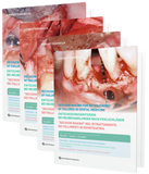
| Naslov | Decision making for retreatment of failures in dental medicine - DVD Compendium |
| Autor | Tabanella, Giorgio |
| Vrsta | Endodontics |
| Datum objave | Sep 21, 2017 |
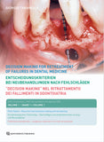
Tabanella, Giorgio
Decision making for retreatment of failures in dental medicine
DVD Compendium
1st Edition 2018
Video DVD
4 DVDs and detailed Engl. booklets in a hardcover, NTSC/PAL
Languages: English, German, Italian
Subject: Oral Surgery
Title-No.: 3800
ISBN 978-1-78698-006-9
Quintessence Publishing, Germany
Increasingly, modern periodontology and implantology face mistreatments, complications, failures, and even iatrogenic damages. It is therefore deemed valuable for colleagues and patients to share protocols, techniques, instruments, materials, and innovations developed to retreat these clinical cases with no room for error. All clinical cases described have only one parameter in common: a medical mistake.
Retreating these "special patients" requires passion, ethics, and the willpower to help patients who present with very high expectations and an exacting psychological makeup. Variables such as periodontal biotype, biologic and anatomic potential for tissue regeneration, and the nature of the underlying osseous architecture surrounding the ailing teeth or endosseous implants will influence the clinical decision making toward achieving optimal "biomimetic" results. The decision to perform implant treatment is ideally driven by the latest evidence-based information, in addition to the educational background, clinical experience, and access to technology of the periodontist; patient and site risk assessment; predictors of disease; economic aspects; probabilities and uncertainties of treatment outcome; and long-term stability of the results.
Volume 1: Endo failure - Tooth loss and perforation of the buccal plate
Case description: A 45-year-old female patient presented with pain and no suppuration on the inferior border of the zygomatic right bone. Clinical examination and three-dimensional (3D) cone beam computed tomography (CBCT) revealed the presence of an endodontic lesion associated with a fenestration of the buccal bone. Simultaneous extraction, bone augmentation, implant insertion, and immediate loading were performed in one surgical phase to reduce the healing period. Only 4 months post-loading, the case was finalized after the pins that were visible through the mucosa were removed. Only two 1 mm incisions were performed to remove the pins without raising a new flap. The pins inserted next to the zygomatic bone were not removed, thus allowing for a minimally invasive surgical approach.
DVD-Video in hardcover; ISBN 978-1-78698-007-6; Title no. 3810; Runtime: 20 min.
Volume 2: Diagnostic failure - Mistreated trauma associated with periapical chronic lesions and anatomic bone deformities
Case description: A 57-year-old male patient presented with a discolored right maxillary central incisor due to a trauma he received about 20 years earlier. He reported that the tooth had been endodontically treated at the time. Since then, neither orthodontic nor prosthetic treatment had been performed. The patient's chief wish was to improve esthetics, although he had been told that not much could be done. The periapical radiograph, followed by a three-dimensional (3D) reconstruction, revealed the presence of an endodontic lesion, an atrophic basal bone, and an unusually large nasopalatine canal. Implant therapy was not an option; however, an advanced bone reconstruction was necessary prior to tooth extraction to avoid the complete collapse of the ridge in the esthetic area. A novel approach performing advanced tissue grafting to speed up the healing process is presented.
2 DVD-Videos in hardcover; ISBN 978-1-78698-008-3; Title no. 3820; Runtime: 48 min.
Volume 3: Perio failure - Recurrent and excessive scaling and root planing
Case description: A 78-year-old male patient presented with a crown fracture of a mandibular central incisor. The clinical examination and radiographic analysis showed the presence of multiple teeth with deep abrasions on the mesial and distal aspects. The patient reported that he had undergone nonsurgical periodontal therapy every 3 months as a supportive therapy, after having received periodontal surgery more than 20 years before. The periapical radiograph of the mandibular incisor showed a chronic periapical lesion due to the necrotic pulp, which was traumatically induced by recurring scaling and root planing. The endodontic lesion perforated the buccal plate so that an implant placement with a simultaneous guided bone regeneration (GBR) procedure was necessary.
DVD-Video in hardcover; ISBN 978-1-78698-009-0; Title no. 3830; Runtime: 19 min.
Volume 4: Iatrogenic bone grafting and implant placement - Ailing dental implants associated with oroantral communication
Case description: A 41-year-old female patient had undergone an implant treatment about 2 years previously. She presented with purulent exudate coming from the right nostril and from one of the dental implants placed after a maxillary sinus elevation. An endodontic lesion was also present next to the infected implant. The implant was mobile and painful. The treatment comprised only two surgical phases: the first phase aimed to eradicate the infection by removing the infected implant and endodontically treated tooth and closing the oroantral communication; the second phase was based on simultaneous implant removal, sinus elevation, simultaneous implant placement, guided bone regeneration (GBR), and immediate loading to reduce the healing time. This clinical case shows the potential for tissue regeneration even after iatrogenic implant dentistry.
2 DVD-Videos in hardcover; ISBN 978-1-78698-010-6; Title no. 3840; Runtime: 39 min.
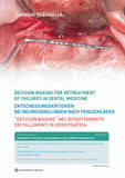
Decision making for retreatment of failures in dental medicine
DVD Compendium
1st Edition 2018
Video DVD
4 DVDs and detailed Engl. booklets in a hardcover, NTSC/PAL
Languages: English, German, Italian
Subject: Oral Surgery
Title-No.: 3800
ISBN 978-1-78698-006-9
Quintessence Publishing, Germany
Increasingly, modern periodontology and implantology face mistreatments, complications, failures, and even iatrogenic damages. It is therefore deemed valuable for colleagues and patients to share protocols, techniques, instruments, materials, and innovations developed to retreat these clinical cases with no room for error. All clinical cases described have only one parameter in common: a medical mistake.
Retreating these "special patients" requires passion, ethics, and the willpower to help patients who present with very high expectations and an exacting psychological makeup. Variables such as periodontal biotype, biologic and anatomic potential for tissue regeneration, and the nature of the underlying osseous architecture surrounding the ailing teeth or endosseous implants will influence the clinical decision making toward achieving optimal "biomimetic" results. The decision to perform implant treatment is ideally driven by the latest evidence-based information, in addition to the educational background, clinical experience, and access to technology of the periodontist; patient and site risk assessment; predictors of disease; economic aspects; probabilities and uncertainties of treatment outcome; and long-term stability of the results.
Volume 1: Endo failure - Tooth loss and perforation of the buccal plate
Case description: A 45-year-old female patient presented with pain and no suppuration on the inferior border of the zygomatic right bone. Clinical examination and three-dimensional (3D) cone beam computed tomography (CBCT) revealed the presence of an endodontic lesion associated with a fenestration of the buccal bone. Simultaneous extraction, bone augmentation, implant insertion, and immediate loading were performed in one surgical phase to reduce the healing period. Only 4 months post-loading, the case was finalized after the pins that were visible through the mucosa were removed. Only two 1 mm incisions were performed to remove the pins without raising a new flap. The pins inserted next to the zygomatic bone were not removed, thus allowing for a minimally invasive surgical approach.
DVD-Video in hardcover; ISBN 978-1-78698-007-6; Title no. 3810; Runtime: 20 min.
Volume 2: Diagnostic failure - Mistreated trauma associated with periapical chronic lesions and anatomic bone deformities
Case description: A 57-year-old male patient presented with a discolored right maxillary central incisor due to a trauma he received about 20 years earlier. He reported that the tooth had been endodontically treated at the time. Since then, neither orthodontic nor prosthetic treatment had been performed. The patient's chief wish was to improve esthetics, although he had been told that not much could be done. The periapical radiograph, followed by a three-dimensional (3D) reconstruction, revealed the presence of an endodontic lesion, an atrophic basal bone, and an unusually large nasopalatine canal. Implant therapy was not an option; however, an advanced bone reconstruction was necessary prior to tooth extraction to avoid the complete collapse of the ridge in the esthetic area. A novel approach performing advanced tissue grafting to speed up the healing process is presented.
2 DVD-Videos in hardcover; ISBN 978-1-78698-008-3; Title no. 3820; Runtime: 48 min.
Volume 3: Perio failure - Recurrent and excessive scaling and root planing
Case description: A 78-year-old male patient presented with a crown fracture of a mandibular central incisor. The clinical examination and radiographic analysis showed the presence of multiple teeth with deep abrasions on the mesial and distal aspects. The patient reported that he had undergone nonsurgical periodontal therapy every 3 months as a supportive therapy, after having received periodontal surgery more than 20 years before. The periapical radiograph of the mandibular incisor showed a chronic periapical lesion due to the necrotic pulp, which was traumatically induced by recurring scaling and root planing. The endodontic lesion perforated the buccal plate so that an implant placement with a simultaneous guided bone regeneration (GBR) procedure was necessary.
DVD-Video in hardcover; ISBN 978-1-78698-009-0; Title no. 3830; Runtime: 19 min.
Volume 4: Iatrogenic bone grafting and implant placement - Ailing dental implants associated with oroantral communication
Case description: A 41-year-old female patient had undergone an implant treatment about 2 years previously. She presented with purulent exudate coming from the right nostril and from one of the dental implants placed after a maxillary sinus elevation. An endodontic lesion was also present next to the infected implant. The implant was mobile and painful. The treatment comprised only two surgical phases: the first phase aimed to eradicate the infection by removing the infected implant and endodontically treated tooth and closing the oroantral communication; the second phase was based on simultaneous implant removal, sinus elevation, simultaneous implant placement, guided bone regeneration (GBR), and immediate loading to reduce the healing time. This clinical case shows the potential for tissue regeneration even after iatrogenic implant dentistry.
2 DVD-Videos in hardcover; ISBN 978-1-78698-010-6; Title no. 3840; Runtime: 39 min.