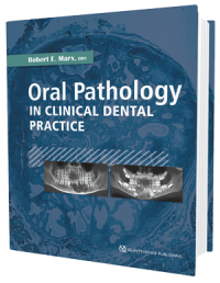
Oral Pathology in Clinical Dental Practice
Marx, Robert E.
Oral Pathology in Clinical Dental Practice
1st edition 2017
Book
Hardcover, 21,6 x 28 cm, 376 pages, 425 images
Language: English
Subjects: Oral/Maxillofacial Surgery, Oral Surgery
Title-No.: 21031
ISBN 978-0-86715-764-2
Quintessence Publishing, USA
Price: 98.00 €
While most dentists do not perform their own histologic testing, all dentists must be able to recognize conditions that may require biopsy or further treatment outside the dentist office. This book does not pretend to be an exhaustive resource on oral pathology; instead, it seeks to provide the practicing clinician with enough information to help identify or at least narrow down the differential for every common lesion or oral manifestation of disease seen in daily practice as well as what to do about them. Organized by type of lesion, mass, or disease, each pathologic entity presented includes the nature of the disease; its predilections, clinical features, radiographic presentation, differential diagnosis, and microscopic features; and the suggested course of action for the dental practitioner as well as the standard treatment regimen. In keeping with the concise nature of the text, all but the rarest disease entities include at least one photograph to illustrate the clinical condition. This book distills the comprehensive information from Dr Marx and Dr Diane Stern's award-winning pathology reference text (Oral and Maxillofacial Pathology: A Rationale for Diagnosis and Treatment, ed 2 [Quintessence, 2012]) into practical guidelines for restorative and general dentists everywhere.
Contents
Chapter 01. Recognizing Abnormalities and Pathologic Conditions
Chapter 02. Red and White Lesions
Chapter 03. Masses Within the Soft Tissues of Oral Mucosa
Chapter 04. Oral Mucosal, Facial, and Neck Masses
Chapter 05. Infectious Diseases of the Jaws and Oral Cavity
Chapter 06. Radiolucent Lesions
Chapter 07. Radiopaque and Mixed Radiolucent-Radiopaque Lesions
Chapter 08. Radiopaque Lesions in Soft Tissue
Chapter 09. Pigmented Lesions of Mucosa and Facial Skin
Chapter 10. Lesions of the Facial Skin and Oral Mucosa
Chapter 11. Exposed Bone Pathologies
Chapter 12. Vascular Lesions of Oral Mucosa and Skin
Robert E. Marx, DDS, professor of surgery and chief of the Division of Oral and Maxillofacial Surgery at the University of Miami Miller School of Medicine, is well known as an educator, researcher, and innovative surgeon. He has pioneered new concepts and treatments for pathologies of the oral and maxillofacial area as well as new techniques in reconstructive surgery. As a researcher, he has made valuable contributions in the use of hyperbaric oxygen following radiation therapy, in the development of platelet-rich plasma, and in elucidating the relationships between smoking and carcinogenesis. He also pioneered the clinical applications of recombinant human bone morphogenetic protein and stem cell use and was the first to identify what is now known worldwide as bisphosphonate induced osteonecrosis of the jaws. For the past 34 years, he has overseen the training of scores of residents and fellows, many of whom have themselves established distinguished careers. His many prestigious awards, including the Harry S. Archer Award, the William J. Gies Award, the Paul Bert Award, and the Donald B. Osbon Award, attest to his dedication and commitment to the field of oral and maxillofacial surgery.
| Naslov | Oral Pathology in Clinical Dental Practice |
| Autor | Marx, Robert E. |
| Vrsta | Oral Pathology |
| Datum objave | Nov 14, 2017 |
Marx, Robert E.
Oral Pathology in Clinical Dental Practice
1st edition 2017
Book
Hardcover, 21,6 x 28 cm, 376 pages, 425 images
Language: English
Subjects: Oral/Maxillofacial Surgery, Oral Surgery
Title-No.: 21031
ISBN 978-0-86715-764-2
Quintessence Publishing, USA
Price: 98.00 €
While most dentists do not perform their own histologic testing, all dentists must be able to recognize conditions that may require biopsy or further treatment outside the dentist office. This book does not pretend to be an exhaustive resource on oral pathology; instead, it seeks to provide the practicing clinician with enough information to help identify or at least narrow down the differential for every common lesion or oral manifestation of disease seen in daily practice as well as what to do about them. Organized by type of lesion, mass, or disease, each pathologic entity presented includes the nature of the disease; its predilections, clinical features, radiographic presentation, differential diagnosis, and microscopic features; and the suggested course of action for the dental practitioner as well as the standard treatment regimen. In keeping with the concise nature of the text, all but the rarest disease entities include at least one photograph to illustrate the clinical condition. This book distills the comprehensive information from Dr Marx and Dr Diane Stern's award-winning pathology reference text (Oral and Maxillofacial Pathology: A Rationale for Diagnosis and Treatment, ed 2 [Quintessence, 2012]) into practical guidelines for restorative and general dentists everywhere.
Contents
Chapter 01. Recognizing Abnormalities and Pathologic Conditions
Chapter 02. Red and White Lesions
Chapter 03. Masses Within the Soft Tissues of Oral Mucosa
Chapter 04. Oral Mucosal, Facial, and Neck Masses
Chapter 05. Infectious Diseases of the Jaws and Oral Cavity
Chapter 06. Radiolucent Lesions
Chapter 07. Radiopaque and Mixed Radiolucent-Radiopaque Lesions
Chapter 08. Radiopaque Lesions in Soft Tissue
Chapter 09. Pigmented Lesions of Mucosa and Facial Skin
Chapter 10. Lesions of the Facial Skin and Oral Mucosa
Chapter 11. Exposed Bone Pathologies
Chapter 12. Vascular Lesions of Oral Mucosa and Skin
Robert E. Marx, DDS, professor of surgery and chief of the Division of Oral and Maxillofacial Surgery at the University of Miami Miller School of Medicine, is well known as an educator, researcher, and innovative surgeon. He has pioneered new concepts and treatments for pathologies of the oral and maxillofacial area as well as new techniques in reconstructive surgery. As a researcher, he has made valuable contributions in the use of hyperbaric oxygen following radiation therapy, in the development of platelet-rich plasma, and in elucidating the relationships between smoking and carcinogenesis. He also pioneered the clinical applications of recombinant human bone morphogenetic protein and stem cell use and was the first to identify what is now known worldwide as bisphosphonate induced osteonecrosis of the jaws. For the past 34 years, he has overseen the training of scores of residents and fellows, many of whom have themselves established distinguished careers. His many prestigious awards, including the Harry S. Archer Award, the William J. Gies Award, the Paul Bert Award, and the Donald B. Osbon Award, attest to his dedication and commitment to the field of oral and maxillofacial surgery.


